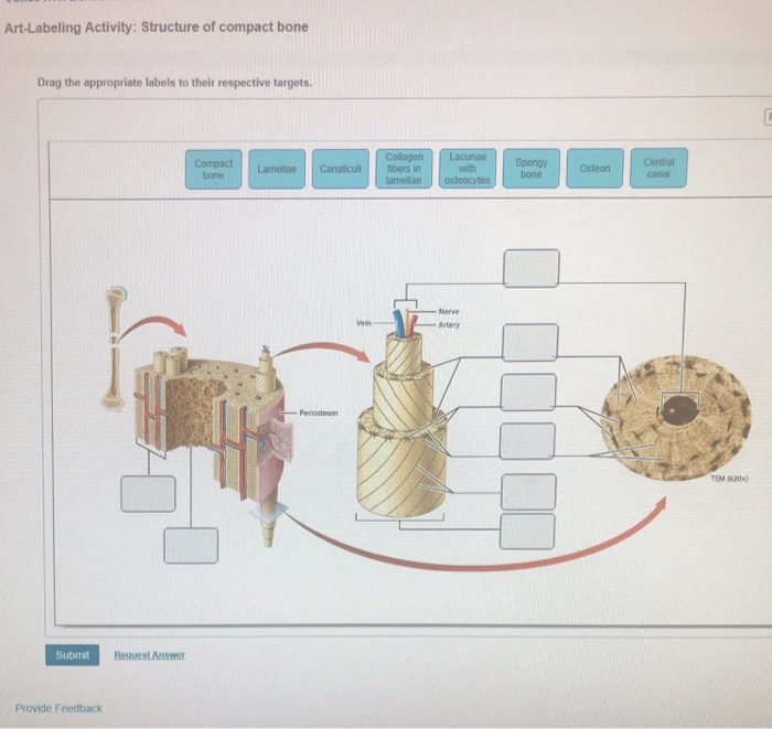
Compact Bone Diagram Labeled - Chapter 6 Page 7 Histologyolm : Skull, clavicle, mandible, scapula, thorax, sternum, humerus, ulna, radius, carpus, phalanges (fingers), metacarpus, spine, pelvis, sacrum, femur, tibia, fibula, tarsus.. Bone diagram creative images compact bone spongy bone and other bone components human anatomy human skeleton labeled back view study anatomy anatomy Begin by identifying the concentric rings of lamellar bone that surround a haversian canal. *free* shipping on qualifying offers. Bones protect the various organs of the body, produce red and white blood cells, store minerals. The osteon consists of a central canal called the osteonic (haversian) canal, which is surrounded by concentric rings (lamellae) of matrix.
In this lab you can explore the bones of the human skeleton using our skeleton viewer that can also be. Corn kernels (primary) early math. Frontal skeleton orthopedic anatomy system publishing, castlecomer on amazon.com. Skull, clavicle, mandible, scapula, thorax, sternum, humerus, ulna, radius, carpus, phalanges (fingers), metacarpus, spine, pelvis, sacrum, femur, tibia, fibula, tarsus. Bones make up the human skeletal system and are used to support the body and help a diagram of the anatomy of a bone, showing the compact bone.
Diagram Diagram Of Bone With Labels Full Version Hd Quality With Labels Coastdiagramleg Pulicentertecno It from image.shutterstock.com Which of the labeled structures in the diagram are fragments of older osteons that have been partially destroyed during bone rebuilding or growth? What is the difference between compact and spongy bone? The remainder is spongelike cancellous bone. Human skeletal diagram labeled bones college ruled composition notebook: Human compact bone is composed of parallel columns made up of concentric bony layers called lamellae organized around channels containing blood vessels, lymph vessels and nerves. A bone is a rigid tissue that constitutes part of the vertebrate skeleton in animals. Long bones, like the tibia and fibula, are those bones whose length exceeds their. Virtual bone labwe need our bones to walk, run, jump and move, but this is not all they do.
There is a printable worksheet available for download here so you can take the quiz with pen and paper.
Compact bone, also called cortical bone, dense bone in which the bony matrix is solidly filled with organic ground substance and inorganic salts, leaving only tiny spaces (lacunae) that contain the related posts of compact bone diagram labeled human cellular respiration diagram. The bones mentioned in each human skeleton chart are: The basic units of compact bone are called osteons or haversian systems. Between the rings of matrix, the bone cells (osteocytes) are located in spaces called lacunae. Corn kernels (primary) early math. Lower jaw (mandible) collar bone. Study guide for students and teachers. What is the difference between compact and spongy bone? 13 photos of the compact bone diagram labeled. There is a printable worksheet available for download here so you can take the quiz with pen and paper. Your bones are strong enough to support your weight, but light enough to allow movement. The remainder is spongelike cancellous bone. Human skeletal diagram labeled bones college ruled composition notebook:
We discuss their function, the different types of bones in the human body long bones: Study guide for students and teachers. The basic units of compact bone are called osteons or haversian systems. What is the difference between compact and spongy bone? Virtual bone labwe need our bones to walk, run, jump and move, but this is not all they do.
Solved Art Labeling Activity Structure Of Compact Bone D Chegg Com from media.cheggcdn.com Cheek bone (zygoma) upper jaw (maxilla). Compact bone, also called cortical bone, is the hard, stiff, smooth, thin, white bone tissue that surrounds all bones in the human body. Which of the labeled structures in the diagram are fragments of older osteons that have been partially destroyed during bone rebuilding or growth? It contains few spaces and provides the spaces between the trabeculae of some spongy bones are filled with red bone marrow. Lower jaw (mandible) collar bone. Blood vessels from the periosteum (see diagram. They protect your delicate internal organs and act as a storehouse for minerals, such as calcium. In this lab you can explore the bones of the human skeleton using our skeleton viewer that can also be.
Bones make up the human skeletal system and are used to support the body and help a diagram of the anatomy of a bone, showing the compact bone.
Long bones, like the tibia and fibula, are those bones whose length exceeds their. Human compact bone is composed of parallel columns made up of concentric bony layers called lamellae organized around channels containing blood vessels, lymph vessels and nerves. Frontal skeleton orthopedic anatomy system publishing, castlecomer on amazon.com. Compact bone, dense bone in which the bony matrix is solidly filled with organic ground substance and inorganic salts, leaving only tiny spaces that contain the osteocytes, or bone cells. The bones mentioned in each human skeleton chart are: Study guide for students and teachers. Compact bone and spongy bone: Lower jaw (mandible) collar bone. Your bones are strong enough to support your weight, but light enough to allow movement. These are mostly compacted bone with little marrow and include most of the bones in flat bones: What is the difference between compact and spongy bone? Cheek bone (zygoma) upper jaw (maxilla). Cancellous bones, compact bone, cortical bone, diaphyses, haversian canal, lamella, marrow cavity, osseous tissue, osteons.
Compact bone consists of outer and inner sheets of lamellar bone (not seen here) and haversian systems, shown here, that run parallel to the long axis of bones. It contains few spaces and provides the spaces between the trabeculae of some spongy bones are filled with red bone marrow. Blood vessels from the periosteum (see diagram. Cheek bone (zygoma) upper jaw (maxilla). Advocate for personal, family, and community health.
Compact Bone Labeling Diagram Quizlet from o.quizlet.com 13 photos of the compact bone diagram labeled. Virtual bone labwe need our bones to walk, run, jump and move, but this is not all they do. There is a printable worksheet available for download here so you can take the quiz with pen and paper. What is the difference between compact and spongy bone? Frontal skeleton orthopedic anatomy system publishing, castlecomer on amazon.com. Compact bone is found on the outside of most bones in the body. How are osteons in compact bone tissue aligned? Compact bone diagram osteon compact bone ap pinterest anatomy human anatomy and.
Compact bone and spongy bone:
The remainder is spongelike cancellous bone. It contains few spaces and provides the spaces between the trabeculae of some spongy bones are filled with red bone marrow. Bones protect the various organs of the body, produce red and white blood cells, store minerals. What is the difference between compact and spongy bone? Bones are very busy even when you are sleeping at night. Virtual bone labwe need our bones to walk, run, jump and move, but this is not all they do. The osteon consists of a central canal called the osteonic (haversian) canal, which is surrounded by concentric rings (lamellae) of matrix. Which of the labeled structures in the diagram are fragments of older osteons that have been partially destroyed during bone rebuilding or growth? Compact bones are made up of osteons while spongy bones are made up of trabeculae. Usually bones that are thin and curved. Compact bone and spongy bone: Study guide for students and teachers. Bone diagram creative images compact bone spongy bone and other bone components human anatomy human skeleton labeled back view study anatomy anatomy